Pelvic Anatomy Xray - File X Ray Of The Pelvis Of A 10 Year Old Female Case 1 Anteroposterior Jpg Wikimedia Commons / Anatomy of the pelvis the pelvis is a ring of bones situated between the spine and the legs.
byAdmin•
0
Pelvic Anatomy Xray - File X Ray Of The Pelvis Of A 10 Year Old Female Case 1 Anteroposterior Jpg Wikimedia Commons / Anatomy of the pelvis the pelvis is a ring of bones situated between the spine and the legs.. Pelvis x ray anatomy in this image you will find the sacroiliac joint acetabular obturator foramina greater trochanter pubic symphysis femoral heads lesser trochanters in it. Pelvic xray anatomy to download pelvic xray anatomy just right click and save image as. Pelvic floor anatomy is complex and is being unraveled by means of magnetic resonance mr imaging. We think this is the most useful anatomy picture that you need. 6.1a, b) is a bony ring consisting of paired innominate bones, the sacrum and coccyx.
48 adductor longus muscle this muscle is the most. Pelvic floor anatomy is complex and is being unraveled by means of magnetic resonance mr imaging. It includes several structures : In an adult, the innominate bones consist of the fused ilium, ischium, and pubis (figure 1). Anatomy of the pelvis the pelvis is a ring of bones situated between the spine and the legs.
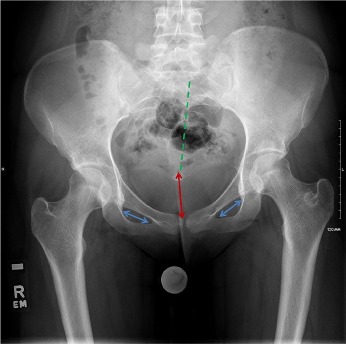
Posterior Anterior Projection Of Pelvic Radiographs In Children Meets The Correct Positioning Criteria Iranian Journal Of Radiology Full Text from neoscriber.org The superior ramus fracture entered the right acetabulum. Anatomy of the pelvis the pelvis is a ring of bones situated between the spine and the legs. Pelvis x ray anatomy in this image you will find the sacroiliac joint acetabular obturator foramina greater trochanter pubic symphysis femoral heads lesser trochanters in it. It is not meant to be a comprehensive reference of imaging anatomy. Mands thorough break down of this commonly used ed diagnostic the pelvic xr. The muscle originates from the body of the pubis and attaches to the pectineal line and proximal part of the linea aspera of femur. For more anatomy content please follow us and visit our website: It is also not meant to present the range of variation across breeds of the domestic animals.
The bony pelvic girdle consists of the innominate bones bilaterally, and the sacrum and coccyx posteriorly.
The pelvis series examines the main pelvic ring, obturator foramina, sacroiliac joints, symphysis pubis, acetabulum, sacral foramina, and the proximal femur. Anatomy of ilioinguinal and iliohypogastric nerves in relation to trocar placement and low transverse incisions. The bony pelvic girdle consists of the innominate bones bilaterally, and the sacrum and coccyx posteriorly. Representative images of normal pelvic anatomy, with select videos. Pelvis anatomy the pelvis is either the lower part of the trunk of the human body between the abdomen and the thighs. In an adult, the innominate bones consist of the fused ilium, ischium, and pubis (figure 1). The bony pelvis, the pelvic cavity, the pelvic floor, and the perineum. No posterior instability is noted despite the pelvic ring gap. The pelvic diaphragm is composed of the ischiococcygeus muscle and levator ani muscle, the latter of which consists of the iliococcygeus, puborectalis, and pubococcygeus muscles. 48 adductor longus muscle this muscle is the most. The imaging anatomy web site is a basic atlas of normal imaging anatomy of domestic animals. Page 3 of 15 (wow) bony pelvis anatomy radiographs ct protocols pelvic ring fx x Each innominate bone is composed of three parts, which fuse at the acetabulum.
Mri Of The Female Pelvis from www.imaios.com It is also not meant to present the range of variation across breeds of the domestic animals. The muscle originates from the body of the pubis and attaches to the pectineal line and proximal part of the linea aspera of femur. The coccyx, commonly referred to as the tailbone, is the smallest of the pelvic bones, and sits inferiorly to the. The imaging anatomy web site is a basic atlas of normal imaging anatomy of domestic animals. 6.1a, b) is a bony ring consisting of paired innominate bones, the sacrum and coccyx. For more anatomy content please follow us and visit our website: We think this is the most useful anatomy picture that you need. Use the mouse scroll wheel to move the images up and down alternatively use the tiny arrows (>>) on both side of the image to move the images.>>) on both side of the image to move the images.
This is an online quiz called elbow xray anatomy.
This is an online quiz called elbow xray anatomy. Angiography invasive angiography is the gold standard modality for assessing pelvic vasculature 3. Tap on/off image to show/hide findings. Pelvic floor anatomy is complex and is being unraveled by means of magnetic resonance mr imaging. Anatomy of ilioinguinal and iliohypogastric nerves in relation to trocar placement and low transverse incisions. Click image to align with top of page. It is also not meant to present the range of variation across breeds of the domestic animals. The bony pelvic girdle consists of the innominate bones bilaterally, and the sacrum and coccyx posteriorly. Our latest youtube film is ready to run. Use the mouse scroll wheel to move the images up and down alternatively use the tiny arrows (>>) on both side of the image to move the images.>>) on both side of the image to move the images. Pelvic anatomy on mri ashish p. Über 7 millionen englischsprachige bücher. The imaging anatomy web site is a basic atlas of normal imaging anatomy of domestic animals.
6.1a, b) is a bony ring consisting of paired innominate bones, the sacrum and coccyx. Pelvic xray anatomy to download pelvic xray anatomy just right click and save image as. Figure 3a schematics show the anatomy of the female pelvic floor at the level of the pelvic diaphragm (a) and the urogenital diaphragm (b). The muscle originates from the body of the pubis and attaches to the pectineal line and proximal part of the linea aspera of femur. We think this is the most useful anatomy picture that you need.
Assessment Of The Young Adult Hip Joint Using Plain Radiographs Springerlink from media.springernature.com Pelvis x ray anatomy in this image you will find the sacroiliac joint acetabular obturator foramina greater trochanter pubic symphysis femoral heads lesser trochanters in it. Page 3 of 15 (wow) bony pelvis anatomy radiographs ct protocols pelvic ring fx x Pelvic xray anatomy to download pelvic xray anatomy just right click and save image as. Erleben sie zuverlässige pharmazeutische beratung. Figure 3a schematics show the anatomy of the female pelvic floor at the level of the pelvic diaphragm (a) and the urogenital diaphragm (b). Pelvic floor anatomy is complex and is being unraveled by means of magnetic resonance mr imaging. This is an online quiz called elbow xray anatomy. What structure is labeled in this radiograph of the pelvis?
Each innominate bone is composed of three parts, which fuse at the acetabulum.
The innominate bones articulate with each other anteriorly and with the sacrum posteriorly. Pelvis anatomy the pelvis is either the lower part of the trunk of the human body between the abdomen and the thighs. The pelvic diaphragm is composed of the ischiococcygeus muscle and levator ani muscle, the latter of which consists of the iliococcygeus, puborectalis, and pubococcygeus muscles. Figure 3a schematics show the anatomy of the female pelvic floor at the level of the pelvic diaphragm (a) and the urogenital diaphragm (b). 48 adductor longus muscle this muscle is the most. This is an online quiz called elbow xray anatomy. Erleben sie zuverlässige pharmazeutische beratung. It is also not meant to present the range of variation across breeds of the domestic animals. In an adult, the innominate bones consist of the fused ilium, ischium, and pubis (figure 1). Anatomy of ilioinguinal and iliohypogastric nerves in relation to trocar placement and low transverse incisions. Über 7 millionen englischsprachige bücher. What structure is labeled in this radiograph of the pelvis? Your pelvis is made up of three bones, the ilium, ischium, and.
Pelvis anatomy the pelvis is either the lower part of the trunk of the human body between the abdomen and the thighs pelvic anatomy. The anorectal hiatus is the only opening in the pelvic diaphragm.